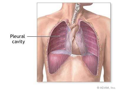
 Figure 11: The pleural cavity (From http://www.nlm.nih.gov/medlineplus/ency/images/ency/fullsize/9749.jpg)
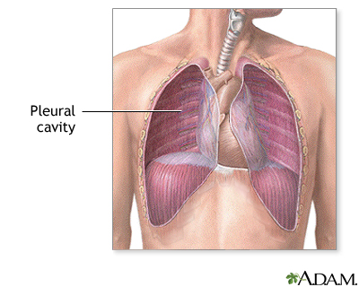
The pleural cavity is the cavity in the thorax which houses the lungs. The lungs and chest wall are covered by the visceral and parietal pleura which are continuous with each other at the hilum. The parietal pleura is attached to the chest wall and the visceral pleura is attached to the lungs [5]. It consists of squamous mesothelium above a thin layer of connective tissue consisting of collagen and some elastic fibers. Between the parietal and visceral pleurae is the pleural space which is filled with a fluid called the pleural fluid [5, 7]. The pleural fluid acts as a lubricant which allows the two pleurae to slide along each other with little friction during inhalation and exhalation [1]. The fluid also provides a certain amount of surface tension which allows the lungs to associate closely with the wall of the chest, which allows for maximum inflation of the alveoli during gas exchange [8].
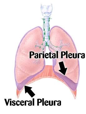
Figure 12: The pleurae which surround the lungs (From http://www.yourlunghealth.org/lung_disease/copd/healthy/graphics/pleura.jpg)