Tuesday, March 25, 2008

Assignment 2: Journal Article Review

C. C. Yen, S. H. Yang, C. Y. Lin, C. M. Chen (2006) Stem cells in the lung parenchyma and prospects for lung injury therapy European Journal of Clinical Investigation 36 (5) , 310–319


Intra-alveolar injury and injury following systemic inflammation of the lungs are the factors which cause acute lung injury which, in its most severe form, results in acute respiratory distress syndrome. This syndrome is a common cause of mortality in hospitals and is currently only managed by treating its underlying causes (for example severe respiratory infection), haemodynamic support, and mechanical ventilation. A new and exciting potential treatment for this syndrome, and many others like it, is being developed in stem cell research. If successful, stem cell manipulation can promote regrowth and healing of damaged lung parenchyma. Research is slow however, as it is hindered by the fact that the mature lung is formed from at least 40 different stem cell lineages, of which each is morphologically differentiated.

Stem cells are cells with a limitless capacity for self-renewal and reinvigorate themselves via mitotic cell division and have the ability to differentiate into any number of cell types. They are known for their plasticity, meaning their ability of the cells to cross cell lineage boundaries. Simply put, a bone marrow stem cell, for example, has the ability to become any type of tissue in the body, from blood to lung tissue. Stem cells are accommodated in niches, or subsets of cells and extra-cellular components that can accommodate stem cells indefinitely and can control their proliferation and renewal.


Figure 1 from Yen et al. (2006) showing the principle of stem cell plasticity.


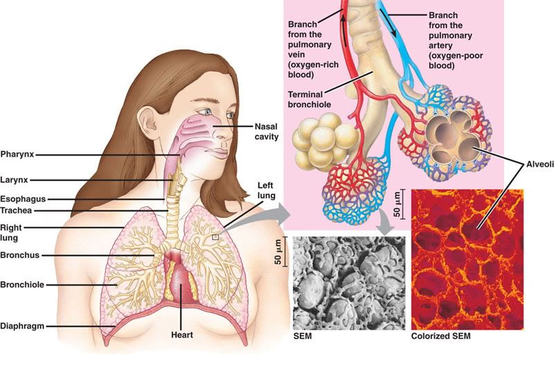
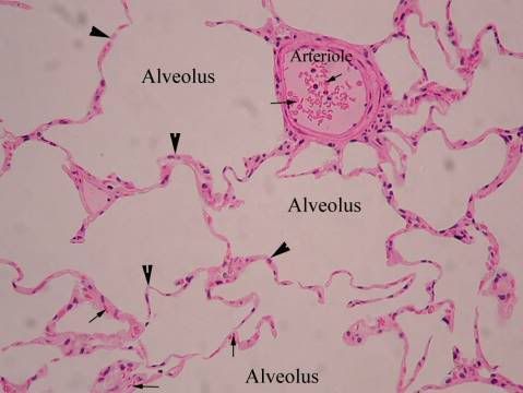
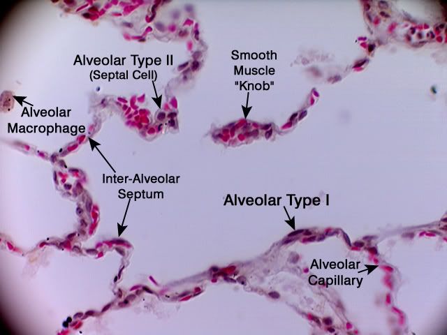
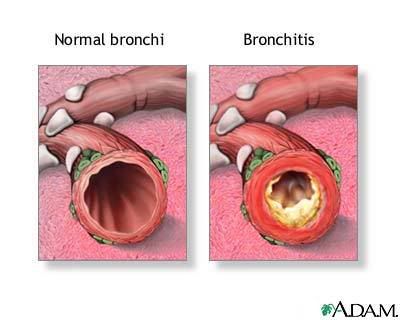
Originally the alveolar type 2 (AT2) cells were thought to be the only stem cells of the lung parenchyma. AT2 cells compose 5% of the surface of the alveoli and are small, polarized epithelial cells which are cuboidal in shape. They have microvilli on the apical surface and organelles typical for eukaryotic cells. They are morphologically distinguished from alveolar type 1 (AT1) cells by the presence of lamellar bodies in the cytoplasm, which secrete the precursors for lung surfactant. Lung surfactants are phospholipids responsible for regulating alveolar surface tension. AT2 have the unique ability to transport sodium from the alveolar space. Water and chloride naturally follow this gradient so the AT2 cells are able to clear fluid from the lungs, which is important in resolving pulmonary edema and inflammatory lung injury.

The original model for stem cell differentiation in the lung parenchyma involved the proliferation and division of the AT2 cells. The AT2 cell was said to give rise to two daughter cells—one of which differentiated into an AT1 cell and the other which remained an AT2 cell. This would typically be stimulated by the loss of either AT1 or AT2 cells. Yen et al. (2006) have brought attention to a significantly modified stem cell model which fits much better with the current knowledge and research on stem cell proliferation. They propose that the AT2 cells maintain in the G0 stage of cell division, meaning they are quiescent. When stimulated by the loss of a nearby AT1 or AT2 cells, they re-enter the cell cycle and divide into two undifferentiated daughter cells which have the potential to become AT1 or AT2 cells. Both AT1 and AT2 cells are capable of cell mediated death (apoptosis) in order to maintain the balance of the epithelial cell population. Exogenous stem cells which are derived from bone marrow and are involved both in later stages of normal lung repair and in repair of serious injury are capable of differentiating into AT1 or AT2 cells depending on the epithelial requirement.


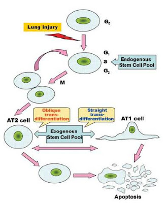
Figure 2 from Yen et al. (2006) showing the resources of stem cells of the alveolar epithelium from endogenous and exogenous pathways.


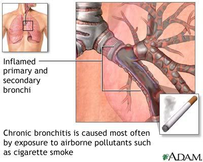
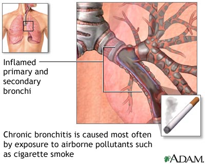
As significant research on stem cells has only been pursued since the 1960s, few molecular signals which regulate stem cells of the lung parenchyma have been discovered. The growth factors KGF and HGF promote the growth, division, and migration of AT2 cells. Retinoic acid (a derivative of vitamin A) has been shown to stimulate the proliferation of AT2 cells as well, including the stimulation of lung repair proceeding injury. It is thought that these effects of retinoic acid are caused by the molecules affecting the genes responsible for lung morphogenesis and increasing recruitment of bone-marrow derived stem cells into the lung.

Stem cell ability to regenerate and repair lung tissue looks to be a promising therapy for injury and disease. Pharmacological therapy could involve using novel growth factors and cytokines discovered to influence lung stem cells to stimulate the body to repair and regenerate itself. In fact, animal experimentation has revealed HGF, KGF, and retinoic acid injections stimulated AT1 and AT2 cell proliferation in the lung. Cellular therapy involves restoring function of damaged tissues via the transplantation of healthy cells. Stem cells have the potential to be transplanted to help restore function to damaged or diseased tissues. Mouse studies already indicate that embryonic stem cells can be used to generate cells which secrete insulin and other hormones, and other studies indicate stem cell transplantation can be used for lung repair. When autologous, stem cell transplantation would overcome the problem of immune rejection. Gene therapy includes the introduction of exogenous genetic material to correct or modify the function of a certain cell. Stem cells may resolve an important problem in gene therapy—the fact that high levels of gene expression without repeated gene transduction cannot be maintained. Genetic modification of stem cells would produce a population of genetically altered cells which would not require repeated procedures.


Figure 3 from Yen et al. (2006) showing prospects for stem cell therapies.


Although significant advances in stem cell research have been made, much more research is required before stem cell therapies become a useful and effective treatment for disease and damage of the lung parenchyma.

This article was exceptionally well written and extensively researched. It is a comprehensive review of relevant information and research on stem cells which can be applied to lung parenchyma injury therapy. Normal lung tissue is described as well as the characteristics of stem cells so the reader can better understand the full implications that stem cells will eventually have on modern medicine—especially disease therapy. The authors introduced a novel and exceptionally in depth model for alveolar epithelium kinetics and induction and proliferation of stem cells in lung (or any) tissue. Unfortunately, I found that the section on stem cell niches and plasticity were far too detailed for a review article, especially since the presence of niches is theoretical in many tissues still. These sections could have been half as long and still detailed enough to fit the flow of the article. Although short, the various therapeutic applications for stem cells the authors describe include both easy to understand explanations and several recent supporting experiments for each therapy. A weakness of this approach, however, is the simple fact that stem cell research is still very basic and the therapies the authors reference are at least a decade away from approaching clinical trials. A problem with the gene therapy section in particular is that it is very general, and only vaguely references lung repair as an application.

Overall this article was easy to read and organized exceptionally well. It is an excellent reference for any scientists or students who wish to enlighten themselves about the potential of stem cells on lung parenchyma repair and regeneration.

Tuesday, March 4, 2008

Assignment 1: The Respiratory System

Figure 1: The Human Respiratory System(from http://kvhs.nbed.nb.ca/gallant/biology/mammalian_respiratory_system.html)

The respiratory system consists of the structures involved in ventilation (the exchange of air between the atmosphere and the lungs) and gas exchange (the exchange of oxygen and carbon dioxide between the lungs and the blood) [5, 7]. This includes the conducting system (the passages which lead from the external atmosphere to the exchange surface of the lungs), the alveoli (the network of sacs that form the surface where oxygen moves from the inhaled air to the blood and carbon dioxide from the blood moves to the air to be exhaled), and the bones and muscles of the chest cavity (the thorax) that assist in ventilation [2, 8]. The respiratory system can be divided into two parts: the upper respiratory tract, consisting of the nasal cavity, pharynx, and larynx, and the lower respiratory tract, consisting of the trachea,two primary bronchi and the bronchioles and alveoli, and the lungs [2].

Fun fact! The entire surface area of the alveoli, if spread out, would cover an entire tennis court-- approximately 70-100 square meters [9].

The Upper Respiratory Tract

Nasal Cavity

Figure 2: Histological preparation of the nasal cavity. (From http://www.visualhistology.com/Visual_Histology_Atlas/VisualHistology_Atlas_2-0-254_1.jpg)


Air is conducted through the nostrils (or nares) of the nose [6]. The first two centimeters of the nares are called the vestibules and are lined with keratinised stratified squamous epithelium. Note the presence of sebaceous glands throughout this epithelium [5, 6]. Hairs line the vestibules and filter matter out of the airstream. The vestibule opens to the nasal cavity (nasopharynx) and the keratinised stratified squamous epithelium transition first to unkeratinized stratified squamous epithelium and then to ciliated pseudostratified columnar epithelium [1]. This epithelium is characteristic for all conductive respiratory surfaces and is thus also called the respiratory epithelium. Goblet cells, which secrete mucus, are present in this epithelium and mucous and serous glands are present in the connective tissue (lamina propria) under this epithelium [8]. These supplement the secretion of the goblet cells. Veins in the lamina propria are in large sinuses called cavernous bodies. The nasal cavity has bony projections called conchae which increase the surface area of the cavity and cause the air passing through to become turbulent. This turbulence allows the nasal cavity to perform its three main functions: filter the air of particulate matter, warm the air before it enters the pharynx, and moisten the air before it reaches the pharynx [1, 2, 8].


Pharynx


The pharynx conducts warm, moist, filtered air from the nasal cavity to the larynx and is divided into three parts [5, 6]. The nasopharynx lies directly behind the nasal cavity and consists of ciliated pseudostratified columnar epithelium [5]. The cilia serve to remove particulate matter from the ventilated air [6]. The oropharynx lies behind the oral cavity and consists of non-keratinized stratified squamous epithelium [5]. The hypopharynx leads into the larynx. The oropharynx and hypopharynx both conduct food as well as air and thus they require the protection against abrasion that non-keratinized stratified squamous epithelium offers [7, 8].


Larynx


Figure 4: The lumen of the larynx. (From http://anatomy.iupui.edu/courses/histo_D502/D502f04/Labs.f04/respiratory%20lab/lary.4x.1.jpg)


The larynx connects the pharynx to the trachea and contains the vocal chords which are used for sound production. The vocal chords are lined with non-keratinized stratified squamous epithelium as well as skeletal muscle in order to control the movement of the vocal folds, which produce sound. Cartilage surrounds the larynx and acts as a support system [7].

The Lower Respiratory Tract

Trachea


Figure 5: The trachea. (From http://www.octc.kctcs.edu/GCaplan/anat2/notes/tra11he.jpg)


The trachea is a tube supported by incomplete rings of cartilage which help to keep it patent, yet allow for stretching of the esophagus for food passage. The trachea connects the larynx to the primary bronchi [2, 6]. It is lined with ciliated pseudostratified columnar epithelium with mucus producing goblet cells and serous cells which secrete an isotonic fluid. The mucus forms a watery layer on top of the epithelium and traps inhaled particles. The cilia beat upwards and carry the debris filled mucus toward the larynx and pharynx where is it swallowed and then digested [1, 8]. The epithelium and lamina propria, consisting of loose connective tissue with elastic fibers, of the trachea are collectively referred to as the mucosa. The layer of connective tissue below the mucosa is referred to as the submucosa, and muco-serous glands in this layer supplement the secretions of the cells in the epithelium. Under the submucosa is the perichondrium [6, 8].


Bronchi and Bronchioles

The trachea divides into the left and right primary bronchi at the carina, which is a ridge in the sagittal plane of the trachea and are characterized by the presence of glands, smooth muscle, and supporting cartilage [1, 2]. They penetrate the lung at an area called the hilum. The primary bronchi undergo extensive branching within each lung, first dividing into lobar bronchi then segmental bronchi, and eventually terminate in the bronchioles, which do not have glands or catrilage and have a thick layer of smooth muscle [2, 8].


Figure 6: One of the bronchi. (From http://umanitoba.ca/faculties/medicine/units/anatomy/images/br22.JPG)


Figure 7: A terminal bronchiole. (From http://www.mc.vanderbilt.edu/histology/labmanual2002/labsection2/Respiratory03_files/image008.jpg)


Larger bronchioles have typical respiratory epithelium-- ciliated pseudostratified columnar epithelium [7]. As the bronchioles decrease in size, the epithelium transitions to ciliated cuboidal cells. Terminal bronchioles contain this ciliated simple cuboidal epithelium and have a thin lamina propria with elastic fibers and smooth muscle cells [1, 8]. They also contain Clara cells which are cilia free dome shaped cells present in the epithelium [8]. They secrete proteins which may assist the bronchi with protection from inflammation. The terminal bronchioles transition into the respiratory bronchioles, which are the first structures which belong to the respiratory portion of the respiratory system. They branch into several alveolar ducts which then lead to the alveoli [1, 8].


The Alveoli


Figure 8: The alveoli (From http://www.octc.kctcs.edu/GCaplan/anat2/notes/image002.jpg)


Alveolar walls (or interalveolar septa) are composed of a thin squamous layer of epithelial cells and a small amounts of collagen, reticular and elastic fibres [1, 8]. Between the septa (in an area known as the interstitium) there is a dense network of pulmonary capillaries which are in direct contact with the epithelium of the alveoli. It is here that gas exchange between the lungs and the blood occurs [1, 2].

The epithelium of the alveoli is composed of two types of cells:

1. Alveolar type I cells. These cells are squamous and can be as thin as 0.05 um. They form 95% of the bulk of the alveolar epithelial surface [2, 8].

2. Alveolar type II cells. These cells are irregular of cuboidal shaped and form small bulges on the alveolar walls. Their cytoplasm contains a large number of granules (called cytosomes) which secrete the precursors of pulmonary surfactant. Surfactant is a mixture of proteins and phospholipids which reduce the surface tension of the alveoli, and prevent their collapse during exhalation, and act as a bactericide [2, 8].


Figure 10: Type I and type II alveolar cells, the interalveolar septum, and the alveolar macrophages. (From http://www.octc.kctcs.edu/GCaplan/anat2/notes/A83_Alveoli_TypeI&II_40X.jpg)

There are no cilia present on the alveolar epithelium and thus no debris which is inhaled can be removed in this fashion [2]. However,another type of cell-- the alveolar macrophage-- migrate freely over the epithelium and ingest any inhaled debris which has escaped the mucus and cilia of the conducting airways [1, 8]. At the end of their lifespan they migrate to the bronchioles and enter the mucus lining to be discharged into the pharynx and swallowed [8].

The alveolar-capillary barrier, which refers to the structures oxygen and carbon dioxide must cross to be exchanged, prevents air bubbles from forming in the blood and prevents blood from entering the lungs. It includes pulmonary surfactant, the alveolar type I cells, the endothelial cells of the capillaries, and the basement membrane between the two [1, 2, 8].

The Pleural Cavity

Figure 11: The pleural cavity (From http://www.nlm.nih.gov/medlineplus/ency/images/ency/fullsize/9749.jpg)


The pleural cavity is the cavity in the thorax which houses the lungs. The lungs and chest wall are covered by the visceral and parietal pleura which are continuous with each other at the hilum. The parietal pleura is attached to the chest wall and the visceral pleura is attached to the lungs [5]. It consists of squamous mesothelium above a thin layer of connective tissue consisting of collagen and some elastic fibers. Between the parietal and visceral pleurae is the pleural space which is filled with a fluid called the pleural fluid [5, 7]. The pleural fluid acts as a lubricant which allows the two pleurae to slide along each other with little friction during inhalation and exhalation [1]. The fluid also provides a certain amount of surface tension which allows the lungs to associate closely with the wall of the chest, which allows for maximum inflation of the alveoli during gas exchange [8].

Figure 12: The pleurae which surround the lungs (From http://www.yourlunghealth.org/lung_disease/copd/healthy/graphics/pleura.jpg)

Respiratory Pathology: Chronic Bronchitis


Figure 13: Chronic bronchitis. (From http://z.about.com/f/p/440/graphics/images/en/17099.jpg)


Chronic bronchitis is a chronic infection and inflammation of the bronchi. This causes a narrowing of the bronchial walls by bronchospasm (contriction of smooth muscle along the bronchi), vasodilation, congestion, and mucosal edema. The continued irritation causes an increase in the number of goblet cells in the bronchi which leads to excessive mucous production. The number of cilia lining the respiratory tract diminish and the excess mucus cause bronchial mucus plugs which in turn cause hyperinflated alveoli. Mucociliary clearance is also reduced which allows for pooling of infectious material which causes further infections and irritation [2].

Symptoms of chronic bronchitis include an expectorating cough (meaning one which produces sputum), shortness of breath, a feeling of tightness in the chest, and wheezing [3, 4].

Chronic bronchitis is diagnosed when an individual has a wet cough for at least three months of the year for two or more years. Pulmonary function testing is performed to reveal obstructed airways, and a chest X-ray is taken to rule out other possible causes of the cough. Blood and sputum samples may also be collected [4].

Bronchodilators and corticosteroids are the primary course of treatment for chronic bronchitis, and antibiotics may be used to treat secondary infections resulting from the disease [4].

One of the most important preemptive measures for preventing chronic bronchitis is to stop smoking, as it significantly increases the risk of this disease [4]. The inhaled cigarette smoke paralyzes the cilia in the respiratory tract and they then cannot sweep the mucus and trapped debris into the esophagus to be digested [4]. Instead the mucus builds and inflames the bronchi. Pathogens trapped in the mucus are not cleared and result in a greater incidence of infection [4, 5].


Figure 14: Smoking as the cause of chronic bronchitis. (From http://www.nlm.nih.gov/medlineplus/ency/images/ency/fullsize/19365.jpg)

References

[1] Junqueira, L.C. and Carneiro, J. 2005. Basic Histology, 11th Edition. McGraw-Hill: Toronto.

[2] Des Jardins, T. 2002. Cardiopulmonary Anatomy and Physiology, 4th Edition. DelmarThompson Learning: Albany, New York.

[3] Wilkins, R.L., Stoller, J.K., and Scanlan, C.L. 2003. Egan's Fundamentals of Respiratory Care, 8th Edition. Mosby: St. Louis, Missouri.

[4] Des Jardins, T. and Burton, G.G. 2006. Clinical Manifestations and Assessment of Respiratory Disease, 5th Edition. Mosby:St. Louis, Missouri.

[5] Respiratory System. Retrieved on March 3, 2008 from http://en.wikipedia.org/wiki/Respiratory_system

[6] The Human Respiratory System. Retrieved on March 3, 2008 from http://users.rcn.com/jkimball.ma.ultranet/BiologyPages/P/Pulmonary.html

[7] Histology Study guide: Respiratory Tract. Retrieved on March 3, 2008 from http://www.siumed.edu/~dking2/crr/rsguide.htm

[8] Volkoff, H. 2008. Biology 3500 Course Notes.

[9] Alveolar Surface Area. Retrieved on March 3, 2008 from http://www.lib.mcg.edu/edu/eshuphysio/program/section4/4ch1/s4ch1_16.htm